N. Saif1,2, H. Hristov1, K. Akiyoshi1, K. Niotis1, I.E. Ariza1, N. Malviya3, P. Lee1, J. Melendez4, G. Sadek5, K. Hackett6, A. Rahman1, J. Meléndez-Cabrero7, C.E. Greer8, L. Mosconi1, R. Krikorian9, R.S. Isaacson1,10
1. Department of Neurology, Weill Cornell Medicine and NewYork-Presbyterian, New York, NY, USA; 2. SUNY Upstate College of Medicine, Syracuse, NY, USA;
3. Albert Einstein College of Medicine, Bronx, NY, USA; 4. Jersey Memory Assessment Service, Health and Community Services, Jersey, United Kingdom; 5. Department of Neurology, Case Western Reserve University & University Hospitals, Cleveland Medical Center, Cleveland, OH, USA; 6. Department of Psychology, Temple University, Philadelphia, PA, USA; 7. Alzheimer’s Prevention Clinic and Research Center to Puerto Rico, San Juan, Puerto Rico, USA; 8. Department of Ophthalmology, Weill Cornell Medicine, New York, NY, USA; 9. Department of Psychiatry & Behavioral Neuroscience, University of Cincinnati College of Medicine, Cincinnati, OH, USA; 10. Center for Brain Health, Alzheimer’s Prevention Clinic, Florida Atlantic University, Boca Raton, FL, USA
Corresponding Author: Richard S. Isaacson, MD, Department of Neurology, Weill Cornell Medicine and NewYork-Presbyterian, 428 E 72nd Street, Suite 400, New York, NY, 10021, USA, Phone: 212-746-4624 Fax: 646-962-0236 Email: rii9004@med.cornell.edu
J Prev Alz Dis 2022;4(9):731-742
Published online April 26, 2022, http://dx.doi.org/10.14283/jpad.2022.44
Abstract
Background: The Comparative Effectiveness Dementia & Alzheimer’s Registry (CEDAR) trial demonstrated that individualized, multi-domain interventions improved cognition and reduced the risk of Alzheimer’s disease (AD). As biological sex is a significant risk factor for AD, it is essential to explore the differential effectiveness of targeted clinical interventions in women vs. men.
Methods: Patients were recruited from an Alzheimer’s Prevention Clinic. Subjects with normal cognition, subjective cognitive decline, or asymptomatic preclinical AD were classified as “Prevention”. Subjects with mild cognitive impairment due to AD or mild AD were classified as “Early Treatment.” The primary outcome was the change from baseline to 18-months on the modified-Alzheimer’s Prevention Cognitive Composite. Secondary outcomes included a cognitive aging composite, AD and cardiovascular (CV) risk scales, and serum biomarkers. Subjects who adhered to >60% of recommendations in the CEDAR trial were included in this a priori sub-group analysis to examine whether individualized intervention effects were modified by sex (n=80).
Results: In the Prevention group, both women (p=0.0205) and men (p=0.0044) demonstrated improvements in cognition with no sex differences (p=0.5244). In the Early Treatment group, there were also no significant sex differences in cognition (p=0.3299). In the Prevention group, women demonstrated greater improvements in the Multi-Ethnic Study of Atherosclerosis risk score (MESA-RS) than men (difference=1.5, p=0.0013). Women in the Early Treatment group demonstrated greater improvements in CV Risk Factors, Aging and Incidence of Dementia (CAIDE) risk score (difference=2.3, p=0.0067), and the MESA-RS (difference=4.1, p<0.001).
Conclusions: Individualized multi-domain interventions are equally effective at improving cognition in women and men. However, personally-tailored interventions led to greater improvements in calculated AD and CV risk, and CV blood biomarkers, in women compared to men. Future study in larger cohorts is necessary to further define sex differences in AD risk reduction in clinical practice.
Key words: Alzheimer’s Prevention Clinic, Alzheimer’s disease; precision medicine, sex-driven differences.
Introduction
Alzheimer’s disease (AD) develops over an extended preclinical period, with amyloid plaques, neurofibrillary tangles, and glucose hypo-metabolism occurring at least 15-20 years before the onset of symptoms (1-4). Considering the limited progress with developing effective, disease-modifying therapeutics to treat AD, along with the fact that an estimated 46 million Americans have preclinical AD, clinical and research efforts have begun to focus on delaying, or possibly preventing, progression to dementia (5).
Research has continued to identify modifiable risk factors throughout the lifespan for AD, such as hypertension, diabetes, and cardiovascular (CV) disease, among others (6-8). Population-attributable risk models have suggested that managing such risk factors can prevent up to one-third of dementia cases, highlighting the immense potential that lies in addressing modifiable risk factors. Several large-scale, randomized controlled trials (RCTs) have shown that multi-domain lifestyle-based interventions, including nutrition, exercise, and cognitive training, can ameliorate vascular and lifestyle-related risk factors, maintain cognitive function, and reduce the risk of dementia (9-12). In an effort to translate these findings into clinical practice, the Comparative Effectiveness Dementia & Alzheimer’s Registry (CEDAR) study showed that individualized clinical management using multi-domain interventions geared for AD risk reduction was feasible in an outpatient clinic setting. Significant improvements in cognition were found from baseline to 18-months when compared with two matched historical control cohorts (National Alzheimer’s Coordinating Center; Rush Memory and Aging Project). Reductions in calculated AD and CV risk scores, and improvements in serum AD risk biomarkers were also found (13).
While it is encouraging that multi-domain interventions may reduce modifiable AD risk, the effectiveness of these approaches has not yet been robustly investigated with consideration for sex. Research has suggested that after increasing age, the most significant risk factor for AD dementia is female sex—two-thirds of AD patients are females (14, 15). Statistical models have indicated that even when accounting for gender-dependent mortality rates, age at death, and differences in lifespan, women still have twice the risk of incidence (16, 17). Furthermore, a growing body of evidence suggests there are stark differences in brain anatomy, function, and age-related morphological changes between men and women (17). Women have been shown to accumulate greater neurofibrillary tangle burden than men when compared at the same amyloid burden level, suggesting an earlier onset of AD (18). One theory posits that estrogen is neuroprotective against AD. As women age, their estrogen levels sharply decline, contributing to the development and progression of AD (19). In men, age-related decline in testosterone levels is associated with increased risk for AD (19). However, men lose testosterone at a significantly slower rate than women lose estrogen (19). Additionally, women traditionally had fewer opportunities for educational attainment and thus have weaker cognitive reserves when compared to men (20).
To our knowledge, the CEDAR study was among the first clinical research trials to evaluate the effectiveness of evidence-based individually-tailored, multi-domain interventions on cognition, AD/CV risk scores, and AD risk biomarkers in real-world clinical practice.
In this sub-analysis, we sought to examine if sex significantly affects the cognitive, CV, and AD risk outcomes in subjects who follow individually-tailored, multi-domain interventions.
Methods
Study Design and Participants
The CEDAR study procedures, baseline population characteristics, and primary results have been previously described in detail (13, 21). In the original study, participants were separated into two groups based on compliance to recommendations: participants who followed >60% of the recommendations were classified as “higher-compliance,” whereas participants who followed ≤60% were classified as “lower-compliance.” In this sub-group analysis, we evaluated the differential effectiveness of the clinical approach itself when considering sex in higher-compliance participants (n=80) from the original study cohort (n=154). Within this cohort, similar to the original study, participants were grouped based on baseline diagnoses: normal cognition, subjective cognitive decline, and preclinical AD participants were classified as “Prevention.” Mild cognitive impairment (MCI) due to AD and mild AD were classified as “Early Treatment.”
All participants were recruited at the Alzheimer’s Prevention Clinic at Weill Cornell Medicine & New-York Presbyterian. Inclusion criteria assessed via an initial telephone screen included a family history of AD and no or minimal cognitive complaints. Exclusion criteria assessed during an in-person evaluation included a diagnosis of moderate-to-severe AD dementia or other dementia; disorders affecting safe engagement in interventions (e.g., malignant disease, major depression, psychotic disorder); or coincident participation in another trial. Participants with a clinical diagnosis of MCI or early mild dementia with negative amyloid neuroimaging were also excluded.
Institutional Review Board approval was obtained on February 16th, 2015, and patients were consented to participate in the Comparative Effectiveness Dementia & Alzheimer’s Registry (Protocol #1408015423). Trial registered at ClinicalTrials.gov (NCT03687710).
Procedures
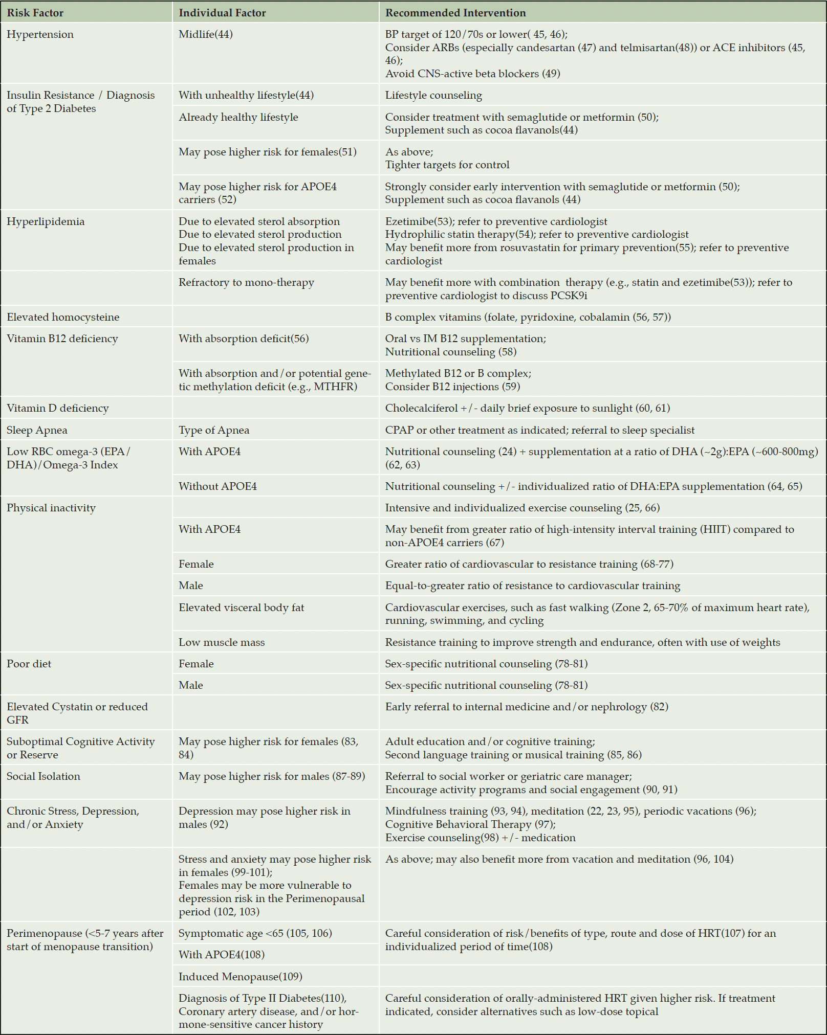
This methodology was outlined in detail in prior publication. In brief, after undergoing baseline clinical assessments, which included a detailed clinical history, physical examination, anthropometrics, blood biomarkers, apolipoprotein-ε4 (APOE-e4) genotyping, and cognitive assessment, patients were given individually-tailored, multi-domain intervention recommendations informed by these clinical and biomarker data. Recommendation categories included patient education/genetic counseling, individualized pharmacological approaches (medications/vitamins/supplements), non-pharmacological approaches (exercise counseling, dietary counseling, vascular risk reduction, sleep hygiene, cognitive engagement, stress reduction, general medical care), and other evidence-based interventions (13, 21). Table 1 outlines which interventions were recommended based on the presence of specific risk factors, as well as differences in various interventions with regards to sex. Additionally, Appendix B in the supplementary material outlines a more detailed application of Table I and our previously published clinical approach (21). It is important to note that these interventions are evidence-based and their clinical application requires a comprehensive evaluation from a qualified healthcare provider. Furthermore, it is necessary for providers to stay current on new research in this field, as evidence continues to emerge on the effectiveness of different interventions across several risk categories, including stress management (e.g., kirtan kriya) (22, 23), nutrition (24), and exercise (25).
Each participant was seen for longitudinal follow-up visits every 6-months, during which continual refinement of interventions would occur. Upon follow-up, each participant was graded as “compliant” or “not compliant” with each recommendation. Afterward, a composite compliance score was calculated as a percentage of recommendations adhered to on a scale of 1–10 (1 represents 0–10% of recommendations, etc.), assessed independently by two clinicians based on patient discussion and patient Likert-scale ratings. Clinicians then collectively assigned an overall compliance score prior to reviewing any follow-up data. Higher-compliance participants were pre-specified as following >60% of all recommendations given, versus lower-compliance participants (≤60%).
Outcomes
The primary outcome was the change in performance on the modified-Alzheimer’s Prevention Initiative Cognitive Composite (m-APCC) from baseline to 18-months. Statistical comparisons were performed between women and men within higher-compliance groups within each diagnostic classification (13, 26).
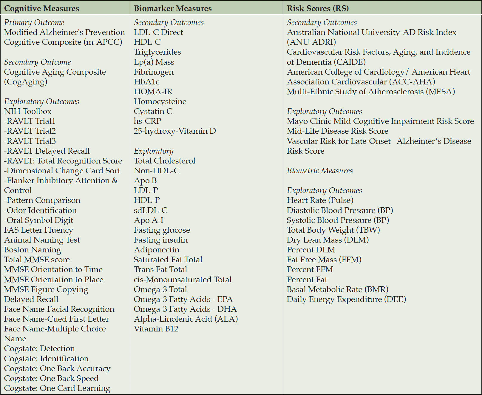
Secondary outcomes included changes on a composite of neuropsychological tests associated with non-pathological cognitive aging (CogAging), two AD risk scales (Australian National University–AD Risk Index (ANU-ADRI), Cardiovascular Risk Factors, Aging and Incidence of Dementia (CAIDE), two CV risk scores (American College of Cardiology/American Heart Association (ACC/AHA), Multi-Ethnic Study of Atherosclerosis (MESA), and AD risk biomarkers (27-30). Exploratory outcomes were chosen a priori based on evidence supporting their use in assessment of AD and/or cardiovascular disease risk. Additionally, when indicated, these measures were used to inform clinical management decisions that target modifiable AD/CVD risk factors. Exploratory outcomes included change in three risk scales, the Mayo Clinic Mild Cognitive Impairment Risk Score (Mayo MCI-RS), Mid-Life Disease Risk Score, and Vascular Risk for Late-Onset Alzheimer’s Disease Risk Score (Vascular Risk for LOAD RS) (31-33).
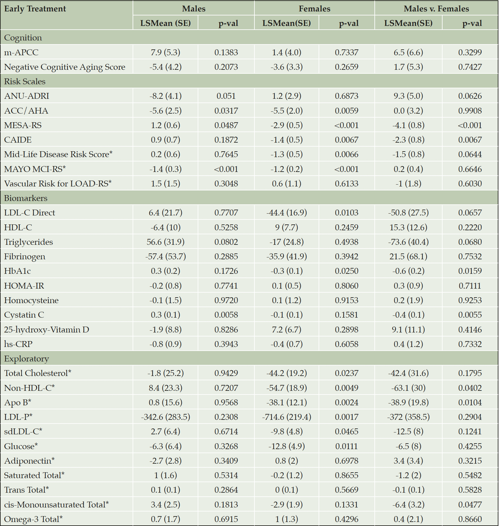
All outcomes are shown in Table 2.
Statistical Methods
General
Participants were grouped based on biological sex and clinical diagnosis. Two-sided P-values were used for all comparisons, with no correction for multiplicity due to the a priori intent to investigate the primary outcome across Diagnosis×Sex groups. All secondary and exploratory analyses may be considered hypothesis-generating and not confirmatory.
Mixed Model Repeated Measures (MMRM)
Change from baseline in all outcomes was analyzed at 18 months for the Full Analysis Set (FAS) using MMRM that included all available data for all participants with at least one follow-up visit. Least squares mean (LSMEAN) estimates at each visit were reported and groups were compared with least squares differences (LSDIFFs). The primary model included diagnostic classification (Prevention/Early Treatment) and sex (male/female) with Diagnosis×Sex interaction, as well as age, baseline score, baseline Mini-Mental State Examination, and visit. The interaction between quantitative compliance and diagnosis group was used to assess whether limiting the analysis to higher-compliance affected the sex sub-groups differently. SAS® V9.4 PROC MIXED was used.
Compliance Adjusted Model
Since participant characteristics may affect compliance levels, predictors of compliance were assessed by fitting a stepwise regression model. To assess the specific impact of compliance, significant baseline predictors of compliance (at α<.05) were identified and corrected for as covariates in the adjusted MMRM, which also included a term for Baseline×Time interaction. LSMEANs estimates from the Diagnosis×Sex interaction using the adjusted model are shown for the primary analysis.
Results
Baseline Demographics
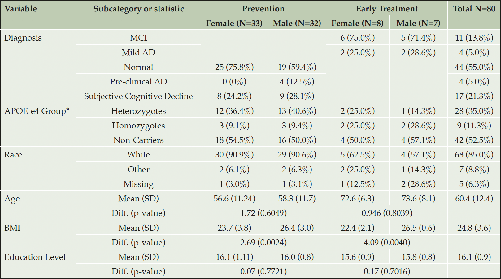
Baseline characteristics are described in Table 3. The mean age of all participants was 60.4 (SD 12.4) years old, with no differences between men and women in the Prevention group (p=0.605) and in the Early Treatment group (p=0.804). There were differences found in body mass index (BMI) between women and men in both the Prevention (p=0.002) and Early Treatment (p=0.004) groups.
Predictors of Compliance
The baseline compliance model identified three baseline parameters that significantly predicted compliance: baseline HbA1c (p<.0001), baseline ACC/AHA risk score (p<.0001), and baseline homocysteine (p=.0225). These parameters were included as co-variates in the Compliance Adjusted Model. The interactive analysis for quantitative compliance and sex resulted in a statistically insignificant interaction (p=0.4518). Each extra point of compliance (complying with an additional 10% of recommendations) resulted in 0.09 point improvement in m-APCC at 18 months within the female group (p=0.7347), and 0.41 points of improvement within the male group (p=0.3133).
Differences found in Prevention Group
Cognition
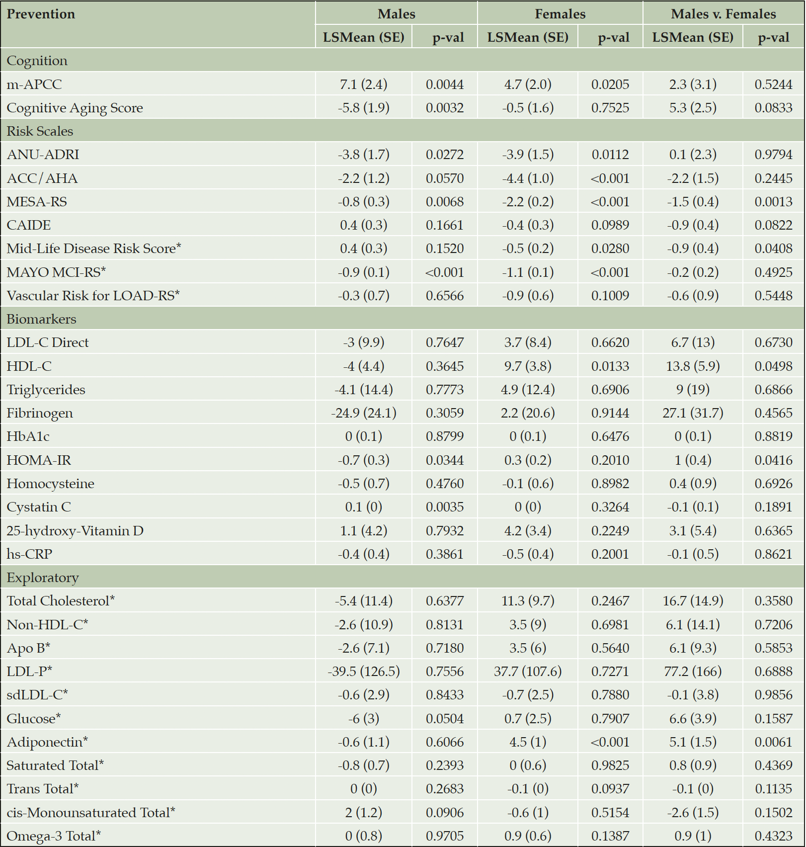
For m-APCC at 18 months, women in the Prevention group improved by 4.7 (2.0) points (p=0.021) and men improved by 7.1 (2.4) points (p=0.004), with no significant difference between these groups (difference= 2.3 [3.1], p=0.524).
For CogAging scores at 18 months, women in the Prevention group improved by 0.5 (1.6) points (p=0.753) and men improved by 5.8 (1.9) points (p=0.003). There was no significant difference between these groups (difference= 5.3 [2.5], p=0.083).
Risk Scales
For ANU-ADRI at 18-months, women in the Prevention group decreased by 3.9 (1.5) points (p=0.011) and men decreased by 3.8 (1.7) points (p=0.027), with no significant difference between these groups (difference= 0.07 [2.3], p=0.979).
For CAIDE at 18 months, women in the Prevention group decreased by 0.4 (0.3) points (p=0.099) and men increased by 0.4 (0.3) points (p=0.166), with no significant difference between these groups (difference= 0.9 [0.4], p=0.082).
For ACC/AHA at 18 months, women in the Prevention group significantly decreased by 4.4 (1.0) points (p=<0.0001) and men decreased by 2.2 (1.2) points (p=0.057), with no significant difference between these groups (difference= 2.2 [1.5], p=0.245).
For MESA at 18 months, women in the Prevention group significantly decreased by 2.2 (0.2) points (p<0.0001) and men significantly decreased by 0.8 (0.3) points (p=0.007), with women significantly improving more than men (difference= 1.5 [0.4], p=0.001).
Biomarkers
In the Prevention group, women showed improvements in HDL-C (9.7 [3.8], p=0.013). Men showed worsening of Cystatin C (0.1 [0.0], p=0.004) and improvement in HOMA-IR ( 0.7 [0.3], p=0.034). Women improved HDL-c more than men (difference= 13.8 [5.9], p=0.050), and men improved HOMA-IR more than women (difference= 1.0 [0.4], p=0.042).
No significant changes were seen within and between women and men for the remaining secondary outcomes (Table IV).
* indicates exploratory outcome measure
Differences found in Early Treatment Group
Cognition
For the m-APCC at 18 months, women in the Early Treatment group improved by 1.4 (4.0) points (p=0.734) and men improved by 7.9 (5.3) points (p=0.138), with no significant difference between the groups (difference= 6.5 [6.6], p=0.330).
For CogAging scores at 18 months, women in the Early Treatment group improved by 3.6 (3.3) points (p=0.266) and men improved by 5.4 (4.2) points (p=0.207), with no significant difference between the groups (difference= 1.7 [5.3], p=0.743).
Risk Scales
For ANU-ADRI at 18 months, women in the Early Treatment group increased by 1.2 (2.9) points (p=0.687) and men decreased by 8.2 (4.1) points (p=0.051), with no significant difference between the groups (difference= 9.3 [5.0], p=0.063).
For CAIDE at 18 months, women in the Early Treatment group decreased by 1.4 (0.5) points (p=0.007) and men increased by 0.9 (0.7) points (p=0.187), with women significantly improving more than men (difference= 2.3 [0.8], p=0.007).
For ACC/AHA at 18 months, women in the Early Treatment group decreased by 5.5 (2.0) points (p=0.006) and men decreased by 5.6 (2.5) points (p=0.032), with no significant difference between the groups (difference= 0.0 [3.2], p=0.991).
For MESA at 18 months, women in the Early Treatment group decreased by 2.9 (0.5) points (p<0.0001) and men decreased by 1.2 (0.6) points (p=0.049), with women significantly improving more than men (difference= 4.1 [0.8], p<0.0001).
Biomarkers
In Early Treatment participants, women showed improvement in HbA1c (-0.3 [0.1], p=0.025), blood glucose (-12.8 [4.9], p=0.011), and LDL-C (-44.4 [16.9] points, p=0.010). Men showed an increase in Cystatin C by (0.3 [0.1], p=0.006). Between women and men, there were differences seen in Cystatin C (difference= 0.4 [0.1], p=0.006) and HbA1c (difference= 0.6 [0.2], p=0.016).
No significant changes were seen within and between women and men for the remaining secondary outcomes (Table V).
* indicates exploratory outcome measure
Discussion
To our knowledge, this is the first study to examine the differential effectiveness of individualized modifiable AD risk reduction between women and men. Previous studies have focused on the role of hormones and sex-specific risk factors when examining differences in AD risk(34), but none have examined if individualized multi-domain interventions result in differences in outcomes related to cognition and AD risk blood biomarkers. While no meaningful differences were seen in the primary outcome of cognitive improvement, we found significant improvements in secondary outcomes including CV risk scales and lipid biomarkers in women compared to men. Further research is needed to draw more definitive conclusions, but our findings suggest that the individualized management approach used by the CEDAR study in a real-world clinic may offer equal cognitive benefits to both women and men, as well as better mitigation of calculated AD and CV risk in women compared to men. Further research with larger cohorts is needed to examine if these better outcomes for women have a significant impact on their risk of developing AD.
In the Prevention group, both women and men improved their m-APCC scores without significant differences between these groups suggesting that an individualized approach that takes sex into consideration may be effective and warranted. Prior studies have suggested that in AD, men demonstrate more rapid rates of cognitive decline than women (35, 36), while other studies support the opposite (37). A recent study suggested that women and men have similar rates of cognitive decline until much later in life when women suffer from a sharp decline (38, 39). Our work highlights the need for larger studies that focus on sex differences in AD-related cognitive trajectories, as the existing body of knowledge lacks conclusive evidence on this issue.
In Early Treatment participants, women improved more than men on both the CAIDE and the MESA. In Prevention participants, women improved more than men on the MESA. The CAIDE is a validated risk index that calculates late-life dementia risk based on midlife vascular risk factors (e.g., body mass index, blood pressure, cholesterol smoking status), while the MESA estimates one’s risk of CV disease incidence over the next ten years using traditional risk factors. These results suggest that women saw greater reductions in their CV risk than men, and later on in the AD spectrum, this reduced CV risk may subsequently reduce the risk of AD. In support of these results, we found that women saw significant improvements in a number of vascular biomarkers, and this may be driving the CV disease risk reduction. Nevertheless, due to the small number of participants in these sub-groups, the significance of these findings are limited. This limitation is further discussed below.
Women in the Prevention group saw significant improvements in HDL cholesterol compared to men. Furthermore, women in the Early Treatment group saw significant improvements in several lipid biomarkers, including total cholesterol, LDL-C, LDL-P, and ApoB. Men in the Early Treatment saw no significant changes in any lipid biomarkers, and the differences between women and men were not found to be significant. However, these findings, when considered alongside the changes found in risk scales, suggest that when patients are further along on the AD spectrum, women may see greater CV benefit than men. It is uncertain why Prevention and Early Treatment women demonstrated improvements in different lipid biomarkers, though differences in age and consequently, estrogen levels may play an important role. Several studies have shown that estrogen can modulate LDL cholesterol absorption, and this relationship will be important to evaluate in further studies (40-42).
Differences found in metabolic markers were more difficult to interpret. Men in the Prevention group saw significantly more improvement in HOMA-IR than Prevention women, while women in the Early Treatment group significantly improved in HbA1c more than Early Treatment men. HOMA-IR and HbA1c have been proven to serve as effective markers for insulin resistance and diabetes diagnoses, which are among the leading risk factors for AD dementia (9). Thus, it is essential that future studies explore the effectiveness of multi-domain interventions targeting glucose metabolism with respect to differences in age, sex, and AD progression.
Our study has several limitations, with a key limitation being that this is a sub-group analysis. Dividing the original sample population into smaller sub-groups resulted in reduced statistical power, as such, detection of significant interactions may have been limited. Due to the small number of participants in the study, specifically in the Early Treatment sub-groups, as well as the vast outcome measures, it is important that our findings are to be considered hypothesis-generating and non-confirmatory. To address this gap, the study authors are in process of expanding a clinical research consortium (currently 6 sites in the United States, 1 in Canada and 1 in the United Kingdom) to expand the cohort, harmonize measures, and yield larger sample sizes upon which more valid conclusions can be made.
An expanded consortium would improve not only the sample size, but also the recruitment of a study population that is more diverse with respect to race, geography, and educational background. Although more than one-third of participants lived outside the New York metropolitan area, our cohort was predominantly Caucasian (85.0%) and, on average, well-educated (16.1 years), and this is an important limitation of our study. The original intent of this study was to have two sites, with the second site (Alzheimer’s Prevention Clinic and Research Center of Puerto Rico) recruiting 100 subjects to begin enrollment in August 2016. Due to a natural disaster (Hurricane Maria, September 2017) that had devastating effects on the clinic, all clinical research operations were halted.
Furthermore, significant differences were found within and between various sub-groups, and we must consider the increased likelihood of false-positive findings due to multiple comparisons across a broad spectrum of outcomes (43). Due to the nature of our analyses, we were not able to determine whether changes in any specific biomarkers were associated with change in cognition in women versus men. With the necessary sample size and power, future studies should seek to perform a Bayesian hierarchical analysis to identify biomarkers that are primarily associated with cognition changes. Nevertheless, future studies must continue to explore differences found in the effectiveness of AD risk reduction strategies within and between sub-groups defined by important baseline characteristics, such as age, sex, and genetic risk factors, as this can expand the body of evidence to inform individualized clinical paradigms.
To investigate sex differences in response to treatment, these analyses only included the participants marked as higher-compliance in the original CEDAR study, and this is an important limitation. This was done to assess for differences in intervention effects within a study cohort that had adhered to a majority of multi-domain interventions recommended. We believe this was the best way to assess for differential response to interventions, as conducting these analyses in lower-compliance participants may have resulted in false-positive findings that were not attributed to following the intervention. Nevertheless, due to the random nature of patient compliance and the inability to randomly assign participants to compliance levels, the sample size is considerably smaller compared to the original CEDAR study. Furthermore, only including higher-compliance participants may introduce bias to our results. We hope to conduct further analyses to continue exploring sex differences within a larger, more diverse sample of participants.
Conclusion
Individualized multi-domain interventions are equally effective in improving cognition in women and men. However, these interventions led to greater AD and CV risk reduction in women compared to men. Future study in larger cohorts with a priori primary and/or secondary outcomes is necessary to examine the effects of sex on AD risk reduction interventions.
Acknowledgements: The authors thank Dorothy Keine, PhD, of 3Prime Medical Writing, LLC for providing medical writing support.
Funding: This study was supported by the Women’s Alzheimer’s Movement; Zuckerman Family Foundation; Ace’s for Alzheimer’s; Altman Family Fund; Harry T. Mangurian, Jr. Foundation; philanthropic support from the patients of the Alzheimer’s Prevention Clinic; NIH/NCATS UL1TR002384 and NIH PO1AG026572. The sponsors had no role in the design and conduct of the study; in the collection, analysis, and interpretation of data; in the preparation of the manuscript; or in the review or approval of the manuscript.
Trial registration: ClinicalTrials.gov NCT03687710.
Conflict of interest: Dr. Isaacson has served as a scientific advisor for Acadia, Biogen, Genentech/Roche, Lilly, and Novo Nordisk. The other authors have nothing to disclose.
Ethical standard: All procedures performed in this study involving human participants were approved by and in accordance with the ethical standards of the IRB. Informed consent was obtained from all participants.
Open Access: This article is distributed under the terms of the Creative Commons Attribution 4.0 International License (http://creativecommons.org/licenses/by/4.0/), which permits use, duplication, adaptation, distribution and reproduction in any medium or format, as long as you give appropriate credit to the original author(s) and the source, provide a link to the Creative Commons license and indicate if changes were made.
References
1. Sperling RA, Aisen PS, Beckett LA, Bennett DA, Craft S, Fagan AM, et al. Toward defining the preclinical stages of Alzheimer’s disease: recommendations from the National Institute on Aging-Alzheimer’s Association workgroups on diagnostic guidelines for Alzheimer’s disease. Alzheimers Dement. 2011;7(3):280-92.
2. Jack CR, Jr., Knopman DS, Jagust WJ, Shaw LM, Aisen PS, Weiner MW, et al. Hypothetical model of dynamic biomarkers of the Alzheimer’s pathological cascade. Lancet Neurol. 2010;9(1):119-28.
3. Jack CR, Jr., Knopman DS, Jagust WJ, Petersen RC, Weiner MW, Aisen PS, et al. Tracking pathophysiological processes in Alzheimer’s disease: an updated hypothetical model of dynamic biomarkers. Lancet Neurol. 2013;12(2):207-16.
4. Jack CR, Bennett DA, Blennow K, Carrillo MC, Dunn B, Haeberlein SB, et al. NIA-AA Research Framework: Toward a biological definition of Alzheimer’s disease. Alzheimer’s & Dementia. 2018;14(4):535-62.
5. Brookmeyer R, Abdalla N, Kawas CH, Corrada MM. Forecasting the prevalence of preclinical and clinical Alzheimer’s disease in the United States. Alzheimers Dement. 2018;14(2):121-9.
6. Gottesman RF, Schneider ALC, Zhou Y, Coresh J, Green E, Gupta N, et al. Association Between Midlife Vascular Risk Factors and Estimated Brain Amyloid Deposition. JAMA. 2017;317(14):1443-50.
7. Janelidze S, Stomrud E, Palmqvist S, Zetterberg H, van Westen D, Jeromin A, et al. Plasma β-amyloid in Alzheimer’s disease and vascular disease. Scientific Reports. 2016;6(1):26801.
8. Edwards III GA, Gamez N, Escobedo Jr. G, Calderon O, Moreno-Gonzalez I. Modifiable Risk Factors for Alzheimer’s Disease. Front Aging Neurosci. 2019;11(146).
9. Livingston G, Sommerlad A, Orgeta V, Costafreda SG, Huntley J, Ames D, et al. Dementia prevention, intervention, and care. Lancet. 2017;390(10113):2673-734.
10. Schelke MW, Attia P, Palenchar DJ, Kaplan B, Mureb M, Ganzer CA, et al. Mechanisms of Risk Reduction in the Clinical Practice of Alzheimer’s Disease Prevention. Front Aging Neurosci. 2018;10:96-.
11. Ngandu T, Lehtisalo J, Solomon A, Levälahti E, Ahtiluoto S, Antikainen R, et al. A 2 year multidomain intervention of diet, exercise, cognitive training, and vascular risk monitoring versus control to prevent cognitive decline in at-risk elderly people (FINGER): a randomised controlled trial. Lancet. 2015;385(9984):2255-63.
12. Kivipelto M, Solomon A, Ahtiluoto S, Ngandu T, Lehtisalo J, Antikainen R, et al. The Finnish Geriatric Intervention Study to Prevent Cognitive Impairment and Disability (FINGER): study design and progress. Alzheimers Dement. 2013;9(6):657-65.
13. Isaacson RS, Hristov H, Saif N, Hackett K, Hendrix S, Melendez J, et al. Individualized clinical management of patients at risk for Alzheimer’s dementia. Alzheimers Dement. 2019;15(12):1588-602.
14. Andrew MK, Tierney MC. The puzzle of sex, gender and Alzheimer’s disease: Why are women more often affected than men? Womens Health (Lond). 2018;14:1745506518817995.
15. Brookmeyer R, Gray S, Kawas C. Projections of Alzheimer’s disease in the United States and the public health impact of delaying disease onset. Am J Public Health. 1998;88(9):1337-42.
16. Viña J, Lloret A. Why women have more Alzheimer’s disease than men: gender and mitochondrial toxicity of amyloid-beta peptide. J Alzheimers Dis. 2010;20 Suppl 2:S527-33.
17. Carter CL, Resnick EM, Mallampalli M, Kalbarczyk A. Sex and gender differences in Alzheimer’s disease: recommendations for future research. J Womens Health (Larchmt). 2012;21(10):1018-23.
18. Buckley RF, Mormino EC, Amariglio RE, Properzi MJ, Rabin JS, Lim YY, et al. Sex, amyloid, and APOE ε4 and risk of cognitive decline in preclinical Alzheimer’s disease: Findings from three well-characterized cohorts. Alzheimers Dement. 2018;14(9):1193-203.
19. Barron AM, Pike CJ. Sex hormones, aging, and Alzheimer’s disease. Front Biosci (Elite Ed). 2012;4:976-97.
20. Scarmeas N, Stern Y. Cognitive reserve: implications for diagnosis and prevention of Alzheimer’s disease. Curr Neurol Neurosci Rep. 2004;4(5):374-80.
21. Isaacson RS, Ganzer CA, Hristov H, Hackett K, Caesar E, Cohen R, et al. The clinical practice of risk reduction for Alzheimer’s disease: A precision medicine approach. Alzheimer’s & dementia : the journal of the Alzheimer’s Association. 2018;14(12):1663-73.
22. Khalsa DS. Stress, Meditation, and Alzheimer’s Disease Prevention: Where The Evidence Stands. Journal of Alzheimer’s Disease. 2015;48:1-12.
23. Khalsa DS, Newberg AB. Spiritual Fitness: A New Dimension in Alzheimer’s Disease Prevention. J Alzheimers Dis. 2021;80(2):505-19.
24. Norwitz NG, Saif N, Ariza IE, Isaacson RS. Precision Nutrition for Alzheimer’s Prevention in ApoE4 Carriers. Nutrients. 2021;13(4):1362.
25. Barha CK, Falck RS, Skou ST, Liu-Ambrose T. Personalising exercise recommendations for healthy cognition and mobility in ageing: time to consider one’s pre-existing function and genotype (Part 2). British Journal of Sports Medicine. 2021;55(6):301.
26. Langbaum JB, Hendrix SB, Ayutyanont N, Chen K, Fleisher AS, Shah RC, et al. An empirically derived composite cognitive test score with improved power to track and evaluate treatments for preclinical Alzheimer’s disease. Alzheimers Dement. 2014;10(6):666-74.
27. Anstey KJ, Cherbuin N, Herath PM. Development of a new method for assessing global risk of Alzheimer’s disease for use in population health approaches to prevention. Prev Sci. 2013;14(4):411-21.
28. Exalto LG, Quesenberry CP, Barnes D, Kivipelto M, Biessels GJ, Whitmer RA. Midlife risk score for the prediction of dementia four decades later. Alzheimers Dement. 2014;10(5):562-70.
29. Muntner P, Colantonio LD, Cushman M, Goff DC, Jr., Howard G, Howard VJ, et al. Validation of the atherosclerotic cardiovascular disease Pooled Cohort risk equations. JAMA. 2014;311(14):1406-15.
30. McClelland RL, Jorgensen NW, Budoff M, Blaha MJ, Post WS, Kronmal RA, et al. 10-Year Coronary Heart Disease Risk Prediction Using Coronary Artery Calcium and Traditional Risk Factors: Derivation in the MESA (Multi-Ethnic Study of Atherosclerosis) With Validation in the HNR (Heinz Nixdorf Recall) Study and the DHS (Dallas Heart Study). J Am Coll Cardiol. 2015;66(15):1643-53.
31. Pankratz VS, Roberts RO, Mielke MM, Knopman DS, Jack CR, Jr., Geda YE, et al. Predicting the risk of mild cognitive impairment in the Mayo Clinic Study of Aging. Neurology. 2015;84(14):1433-42.
32. Kivipelto M, Ngandu T, Laatikainen T, Winblad B, Soininen H, Tuomilehto J. Risk score for the prediction of dementia risk in 20 years among middle aged people: a longitudinal, population-based study. Lancet Neurol. 2006;5(9):735-41.
33. Reitz C, Tang MX, Schupf N, Manly JJ, Mayeux R, Luchsinger JA. A summary risk score for the prediction of Alzheimer disease in elderly persons. Arch Neurol. 2010;67(7):835-41.
34. Rahman A, Jackson H, Hristov H, Isaacson RS, Saif N, Shetty T, et al. Sex and Gender Driven Modifiers of Alzheimer’s: The Role for Estrogenic Control Across Age, Race, Medical, and Lifestyle Risks. Front Aging Neurosci. 2019;11:315-.
35. Maylor EA, Reimers S, Choi J, Collaer ML, Peters M, Silverman I. Gender and Sexual Orientation Differences in Cognition Across Adulthood: Age Is Kinder to Women than to Men Regardless of Sexual Orientation. Archives of Sexual Behavior. 2007;36(2):235-49.
36. Barrett-Connor E, Kritz-Silverstein D. Gender differences in cognitive function with age: the Rancho Bernardo study. J Am Geriatr Soc. 1999;47(2):159-64.
37. Singh-Manoux A, Kivimaki M, Glymour MM, Elbaz A, Berr C, Ebmeier KP, et al. Timing of onset of cognitive decline: results from Whitehall II prospective cohort study. BMJ. 2012;344:d7622.
38. Proust-Lima C, Amieva H, Letenneur L, Orgogozo JM, Jacqmin-Gadda H, Dartigues JF. Gender and education impact on brain aging: a general cognitive factor approach. Psychol Aging. 2008;23(3):608-20.
39. Read S, Pedersen NL, Gatz M, Berg S, Vuoksimaa E, Malmberg B, et al. Sex differences after all those years? Heritability of cognitive abilities in old age. J Gerontol B Psychol Sci Soc Sci. 2006;61(3):P137-43.
40. Iorga A, Cunningham CM, Moazeni S, Ruffenach G, Umar S, Eghbali M. The protective role of estrogen and estrogen receptors in cardiovascular disease and the controversial use of estrogen therapy. Biol Sex Differ. 2017;8(1):33-.
41. Lagranha CJ, Silva TLA, Silva SCA, Braz GRF, da Silva AI, Fernandes MP, et al. Protective effects of estrogen against cardiovascular disease mediated via oxidative stress in the brain. Life Sci. 2018;192:190-8.
42. Mendelsohn ME, Karas RH. The protective effects of estrogen on the cardiovascular system. N Engl J Med. 1999;340(23):1801-11.
43. Wang R, Lagakos SW, Ware JH, Hunter DJ, Drazen JM. Statistics in Medicine — Reporting of Subgroup Analyses in Clinical Trials. New England Journal of Medicine. 2007;357(21):2189-94.
44. Mastroiacovo D, Kwik-Uribe C, Grassi D, Necozione S, Raffaele A, Pistacchio L, et al. Cocoa flavanol consumption improves cognitive function, blood pressure control, and metabolic profile in elderly subjects: the Cocoa, Cognition, and Aging (CoCoA) Study–a randomized controlled trial. Am J Clin Nutr. 2015;101(3):538-48.
45. The SMIftSRG. Effect of Intensive vs Standard Blood Pressure Control on Probable Dementia: A Randomized Clinical Trial. JAMA. 2019;321(6):553-61.
46. Nasrallah IM, Gaussoin SA, Pomponio R, Dolui S, Erus G, Wright CB, et al. Association of Intensive vs Standard Blood Pressure Control With Magnetic Resonance Imaging Biomarkers of Alzheimer Disease: Secondary Analysis of the SPRINT MIND Randomized Trial. JAMA Neurology. 2021;78(5):568-77.
47. Hajjar I, Okafor M, McDaniel D, Obideen M, Dee E, Shokouhi M, et al. Effects of Candesartan vs Lisinopril on Neurocognitive Function in Older Adults With Executive Mild Cognitive Impairment: A Randomized Clinical Trial. JAMA Network Open. 2020;3(8):e2012252-e.
48. Liu C-H, Sung P-S, Li Y-R, Huang W-K, Lee T-W, Huang C-C, et al. Telmisartan use and risk of dementia in type 2 diabetes patients with hypertension: A population-based cohort study. PLOS Medicine. 2021;18(7):e1003707.
49. Holm H, Ricci F, Di Martino G, Bachus E, Nilsson ED, Ballerini P, et al. Beta-blocker therapy and risk of vascular dementia: A population-based prospective study. Vascul Pharmacol. 2020;125-126:106649.
50. Bendlin BB. Antidiabetic therapies and Alzheimer disease. Dialogues Clin Neurosci. 2019;21(1):83-91.
51. Hayden KM, Zandi PP, Lyketsos CG, Khachaturian AS, Bastian LA, Charoonruk G, et al. Vascular risk factors for incident Alzheimer disease and vascular dementia: the Cache County study. Alzheimer Dis Assoc Disord. 2006;20(2):93-100.
52. Matsuzaki T, Sasaki K, Tanizaki Y, Hata J, Fujimi K, Matsui Y, et al. Insulin resistance is associated with the pathology of Alzheimer disease: the Hisayama study. Neurology. 2010;75(9):764-70.
53. Kato ET, Cannon CP, Blazing MA, Bohula E, Guneri S, White JA, et al. Efficacy and Safety of Adding Ezetimibe to Statin Therapy Among Women and Men: Insight From IMPROVE-IT (Improved Reduction of Outcomes: Vytorin Efficacy International Trial). Journal of the American Heart Association.6(11):e006901.
54. Chu C-S, Tseng P-T, Stubbs B, Chen T-Y, Tang C-H, Li D-J, et al. Use of statins and the risk of dementia and mild cognitive impairment: A systematic review and meta-analysis. Scientific Reports. 2018;8(1):5804.
55. Mora S, Glynn RJ, Hsia J, MacFadyen JG, Genest J, Ridker PM. Statins for the Primary Prevention of Cardiovascular Events in Women With Elevated High-Sensitivity C-Reactive Protein or Dyslipidemia. Circulation. 2010;121(9):1069-77.
56. Smith AD, Smith SM, de Jager CA, Whitbread P, Johnston C, Agacinski G, et al. Homocysteine-lowering by B vitamins slows the rate of accelerated brain atrophy in mild cognitive impairment: a randomized controlled trial. PLoS One. 2010;5(9):e12244.
57. Price BR, Wilcock DM, Weekman EM. Hyperhomocysteinemia as a Risk Factor for Vascular Contributions to Cognitive Impairment and Dementia. Front Aging Neurosci. 2018;10(350).
58. Schelke MW, Hackett K, Chen JL, Shih C, Shum J, Montgomery ME, et al. Nutritional interventions for Alzheimer’s prevention: a clinical precision medicine approach. Ann N Y Acad Sci. 2016;1367(1):50-6.
59. Moll S, Varga EA. Homocysteine and MTHFR Mutations. Circulation. 2015;132(1):e6-9.
60. Annweiler C, Llewellyn DJ, Beauchet O. Low serum vitamin D concentrations in Alzheimer’s disease: a systematic review and meta-analysis. J Alzheimers Dis. 2013;33(3):659-74.
61. Annweiler C, Montero-Odasso M, Llewellyn DJ, Richard-Devantoy S, Duque G, Beauchet O. Meta-analysis of memory and executive dysfunctions in relation to vitamin D. J Alzheimers Dis. 2013;37(1):147-71.
62. Yassine HN, Cordova I, He X, Solomon V, Mack WJ, Harrington MG, et al. Refining omega-3 supplementation trials in APOE4 carriers for dementia prevention. Alzheimer’s & Dementia. 2020;16(S5):e039029.
63. Lin P-Y, Cheng C, Satyanarayanan SK, Chiu L-T, Chien Y-C, Chuu C-P, et al. Omega-3 fatty acids and blood-based biomarkers in Alzheimer’s disease and mild cognitive impairment: A randomized placebo-controlled trial. Brain, Behavior, and Immunity. 2022;99:289-98.
64. Macaron T, Giudici KV, Bowman GL, Sinclair A, Stephan E, Vellas B, et al. Associations of Omega-3 fatty acids with brain morphology and volume in cognitively healthy older adults: A narrative review. Ageing Research Reviews. 2021;67:101300.
65. Jernerén F, Elshorbagy AK, Oulhaj A, Smith SM, Refsum H, Smith AD. Brain atrophy in cognitively impaired elderly: the importance of long-chain ω-3 fatty acids and B vitamin status in a randomized controlled trial. Am J Clin Nutr. 2015;102(1):215-21.
66. Smith JC, Nielson KA, Woodard JL, Seidenberg M, Durgerian S, Hazlett KE, et al. Physical activity reduces hippocampal atrophy in elders at genetic risk for Alzheimer’s disease. Front Aging Neurosci. 2014;6:61-.
67. Tokgöz S, Claassen JAHR. Exercise as Potential Therapeutic Target to Modulate Alzheimer’s Disease Pathology in APOE ε4 Carriers: A Systematic Review. Cardiology and Therapy. 2021;10(1):67-88.
68. Baker LD, Frank LL, Foster-Schubert K, Green PS, Wilkinson CW, McTiernan A, et al. Effects of aerobic exercise on mild cognitive impairment: a controlled trial. Arch Neurol. 2010;67(1):71-9.
69. Hörder H, Johansson L, Guo X, Grimby G, Kern S, Östling S, et al. Midlife cardiovascular fitness and dementia: A 44-year longitudinal population study in women. Neurology. 2018;90(15):e1298-e305.
70. Fallah N, Mitnitski A, Middleton L, Rockwood K. Modeling the impact of sex on how exercise is associated with cognitive changes and death in older Canadians. Neuroepidemiology. 2009;33(1):47-54.
71. Clifford A, Hogervorst E, Bandelow S. O1-02-02: Preventing cognitive decline in the elderly through physical activity in midlife. Alzheimer’s & Dementia. 2011;7(4S_Part_3):S95-S6.
72. Eggermont L, Swaab D, Luiten P, Scherder E. Exercise, cognition and Alzheimer’s disease: more is not necessarily better. Neurosci Biobehav Rev. 2006;30(4):562-75.
73. Kwak YS, Um SY, Son TG, Kim DJ. Effect of regular exercise on senile dementia patients. Int J Sports Med. 2008;29(6):471-4.
74. Liu-Ambrose T, Nagamatsu LS, Graf P, Beattie BL, Ashe MC, Handy TC. Resistance training and executive functions: a 12-month randomized controlled trial. Arch Intern Med. 2010;170(2):170-8.
75. Cassilhas RC, Viana VA, Grassmann V, Santos RT, Santos RF, Tufik S, et al. The impact of resistance exercise on the cognitive function of the elderly. Med Sci Sports Exerc. 2007;39(8):1401-7.
76. Best JR, Chiu BK, Liang Hsu C, Nagamatsu LS, Liu-Ambrose T. Long-Term Effects of Resistance Exercise Training on Cognition and Brain Volume in Older Women: Results from a Randomized Controlled Trial. J Int Neuropsychol Soc. 2015;21(10):745-56.
77. Weuve J, Kang JH, Manson JE, Breteler MMB, Ware JH, Grodstein F. Physical Activity, Including Walking, and Cognitive Function in Older Women. JAMA. 2004;292(12):1454-61.
78. Scheyer O, Rahman A, Hristov H, Berkowitz C, Isaacson RS, Diaz Brinton R, et al. Female Sex and Alzheimer’s Risk: The Menopause Connection. J Prev Alzheimers Dis. 2018;5(4):225-30.
79. Nagel G, Altenburg HP, Nieters A, Boffetta P, Linseisen J. Reproductive and dietary determinants of the age at menopause in EPIC-Heidelberg. Maturitas. 2005;52(3-4):337-47.
80. Dorjgochoo T, Kallianpur A, Gao Y-T, Cai H, Yang G, Li H, et al. Dietary and lifestyle predictors of age at natural menopause and reproductive span in the Shanghai Women’s Health Study. Menopause. 2008;15(5):924-33.
81. Dunneram Y, Greenwood DC, Burley VJ, Cade JE. Dietary intake and age at natural menopause: results from the UK Women’s Cohort Study. J Epidemiol Community Health. 2018;72(8):733-40.
82. Seliger SL, Siscovick DS, Stehman-Breen CO, Gillen DL, Fitzpatrick A, Bleyer A, et al. Moderate Renal Impairment and Risk of Dementia among Older Adults: The Cardiovascular Health Cognition Study. Journal of the American Society of Nephrology. 2004;15(7):1904.
83. Letenneur L, Launer LJ, Andersen K, Dewey ME, Ott A, Copeland JR, et al. Education and the risk for Alzheimer’s disease: sex makes a difference. EURODEM pooled analyses. EURODEM Incidence Research Group. Am J Epidemiol. 2000;151(11):1064-71.
84. Flicker L, Almeida O, Acres J, Lê ML, Tuohy R, Jamrozik K, et al. Predictors of impaired cognitive function in men over the age of 80 years: results from the Health in Men Study. Age and ageing. 2005;34:77-80.
85. Olatunji BO, Kauffman BY, Meltzer S, Davis ML, Smits JA, Powers MB. Cognitive-behavioral therapy for hypochondriasis/health anxiety: a meta-analysis of treatment outcome and moderators. Behav Res Ther. 2014;58:65-74.
86. Ihle A, Oris M, Fagot D, Baeriswyl M, Guichard E, Kliegel M. The Association of Leisure Activities in Middle Adulthood with Cognitive Performance in Old Age: The Moderating Role of Educational Level. Gerontology. 2015;61(6):543-50.
87. Crooks VC, Lubben J, Petitti DB, Little D, Chiu V. Social network, cognitive function, and dementia incidence among elderly women. Am J Public Health. 2008;98(7):1221-7.
88. Sutin AR, Stephan Y, Luchetti M, Terracciano A. Loneliness and Risk of Dementia. The Journals of Gerontology: Series B. 2018;75(7):1414-22.
89. Zebhauser A, Hofmann-Xu L, Baumert J, Häfner S, Lacruz ME, Emeny RT, et al. How much does it hurt to be lonely? Mental and physical differences between older men and women in the KORA-Age Study. Int J Geriatr Psychiatry. 2014;29(3):245-52.
90. Kuiper JS, Zuidersma M, Oude Voshaar RC, Zuidema SU, van den Heuvel ER, Stolk RP, et al. Social relationships and risk of dementia: A systematic review and meta-analysis of longitudinal cohort studies. Ageing Res Rev. 2015;22:39-57.
91. Cotterell N, Buffel T, Phillipson C. Preventing social isolation in older people. Maturitas. 2018;113:80-4.
92. Fuhrer R, Dufouil C, Dartigues JF. Exploring sex differences in the relationship between depressive symptoms and dementia incidence: prospective results from the PAQUID Study. J Am Geriatr Soc. 2003;51(8):1055-63.
93. Salmoirago-Blotcher E, Trivedi D, Dunsiger S, Harris K, Breault C, Yang Santos C, et al. Exploring Effects of Aerobic Exercise and Mindfulness Training on Cognitive Function in Older Adults at Risk of Dementia: A Feasibility, Proof-of-Concept Study. American Journal of Alzheimer’s Disease & Other Dementias®. 2021;36:15333175211039094.
94. Ng TKS, Fam J, Feng L, Cheah IK-M, Tan CT-Y, Nur F, et al. Mindfulness improves inflammatory biomarker levels in older adults with mild cognitive impairment: a randomized controlled trial. Translational Psychiatry. 2020;10(1):21.
95. Innes KE, Selfe TK. Meditation as a Therapeutic Intervention for Adults at Risk for Alzheimer’s Disease – Potential Benefits and Underlying Mechanisms. Frontiers in Psychiatry. 2014;5(40).
96. Epel ES, Puterman E, Lin J, Blackburn EH, Lum PY, Beckmann ND, et al. Meditation and vacation effects have an impact on disease-associated molecular phenotypes. Translational Psychiatry. 2016;6(8):e880-e.
97. Reid LD, Avens FE, Walf AA. Cognitive behavioral therapy (CBT) for preventing Alzheimer’s disease. Behavioural Brain Research. 2017;334:163-77.
98. Wegner M, Helmich I, Machado S, Nardi AE, Arias-Carrion O, Budde H. Effects of exercise on anxiety and depression disorders: review of meta- analyses and neurobiological mechanisms. CNS Neurol Disord Drug Targets. 2014;13(6):1002-14.
99. Wilson RS, Begeny CT, Boyle PA, Schneider JA, Bennett DA. Vulnerability to stress, anxiety, and development of dementia in old age. Am J Geriatr Psychiatry. 2011;19(4):327-34.
100. Peavy GM, Lange KL, Salmon DP, Patterson TL, Goldman S, Gamst AC, et al. The effects of prolonged stress and APOE genotype on memory and cortisol in older adults. Biol Psychiatry. 2007;62(5):472-8.
101. Borenstein AR, Copenhaver CI, Mortimer JA. Early-life risk factors for Alzheimer disease. Alzheimer Dis Assoc Disord. 2006;20(1):63-72.
102. Bromberger JT, Kravitz HM, Chang YF, Cyranowski JM, Brown C, Matthews KA. Major depression during and after the menopausal transition: Study of Women’s Health Across the Nation (SWAN). Psychol Med. 2011;41(9):1879-88.
103. Szoeke CEI, Robertson JS, Rowe CC, Yates P, Campbell K, Masters CL, et al. The Women’s Healthy Ageing Project: Fertile ground for investigation of healthy participants ‘at risk’ for dementia. International Review of Psychiatry. 2013;25(6):726-37.
104. Hoge EA, Chen MM, Orr E, Metcalf CA, Fischer LE, Pollack MH, et al. Loving-Kindness Meditation practice associated with longer telomeres in women. Brain, Behavior, and Immunity. 2013;32:159-63.
105. Craig MC, Maki PM, Murphy DG. The Women’s Health Initiative Memory Study: findings and implications for treatment. Lancet Neurol. 2005;4(3):190-4.
106. Yoo JE, Shin DW, Han K, Kim D, Won HS, Lee J, et al. Female reproductive factors and the risk of dementia: a nationwide cohort study. European Journal of Neurology. 2020;27(8):1448-58.
107. LeBlanc ES, Janowsky J, Chan BKS, Nelson HD. Hormone Replacement Therapy and CognitionSystematic Review and Meta-analysis. JAMA. 2001;285(11):1489-99.
108. Zandi PP, Carlson MC, Plassman BL, Welsh-Bohmer KA, Mayer LS, Steffens DC, et al. Hormone replacement therapy and incidence of Alzheimer disease in older women: the Cache County Study. Jama. 2002;288(17):2123-9.
109. Rasgon NL, Geist CL, Kenna HA, Wroolie TE, Williams KE, Silverman DH. Prospective randomized trial to assess effects of continuing hormone therapy on cerebral function in postmenopausal women at risk for dementia. PLoS One. 2014;9(3):e89095.
110. Carcaillon L, Brailly-Tabard S, Ancelin M-L, Rouaud O, Dartigues J-F, Guiochon-Mantel A, et al. High plasma estradiol interacts with diabetes on risk of dementia in older postmenopausal women. Neurology. 2014;82(6):504.

